Temporal expression analysis of angiogenesis-related genes in brain development
Vascular Cell. 2012;
Received: 3 January 2012 | Accepted: 21 August 2012 | Published: 1 October 2012
Vascular Cell ISSN: 2045-824X
Abstract
Background
The current knowledge on molecular pathogenesis of cerebral vascular malformations (CVM), which are believed to arise during development, is very limited. To unravel the molecular mechanisms involved in CVMs, a detailed understanding of the brain vascular development at molecular level is crucial. In this study, we aimed to explore the temporal and comparative expression profile of angiogenesis-related genes in the establishment of brain vasculature.
Methods
Expression of a total of 113 angiogenesis-related genes during murine brain development has been analyzed using low-density array systems designed for angiogenesis-related genes. Bai1 (brain specific angiogenesis inhibitor-1), a recently identified novel anti-angiogenic gene, has been selected for further characterization.
Results
We found that 62 out of 113 analyzed genes have expression in brain development at varying levels. Nineteen of these were differentially expressed between embryonic and postnatal stages (>1.5 fold). Bai1 is strongly expressed on growing blood vessels of cerebral cortex and hippocampus, partially expressed in the lateral regions of striatum, but mostly absent on the thalamus.
Conclusion
By showing the comparative expression analysis of angiogenesis-related genes throughout brain development, the data presented here will be a crucial addition to further functional studies on cerebrovascular research.
Keywords
Cerebrovascular development Arteriovenous malformations Angiogenesis Bai1Introduction
Embryonic vascular development is composed of a highly complex order of events that involve a variety of cell-cell interactions and a tightly balanced regulation of a wide range of functional molecules including growth factors and their receptors, transcription factors, cytokines, chemokines, proteases and their inhibitors, adhesion molecules and numerous matrix proteins. Actions of these factors must be well orchestrated in terms of time, space and dosage to form a functional vascular network [1–3].
In 1986 McCormick pathologically classified cerebrovascular malformations into the four following subgroups: Arteriovenous malformations (AVMs), cavernous malformations, venous angiomas and capillary telengiectasias [4]. Among these, AVMs are considered the most dangerous, with a high risk of intracranial hemorrhage [5]. AVMs are characterized with high-flow tangles of malformed arteries and veins without an intervening capillary bed. They bear a morphological resemblance to vascular plexus seen in development and are generally considered as congenital, resulting from defects in embryonic vascular specialization of the central nervous system [6]. Features of spontaneous hemorrhage, recurrence, growth and regression strongly suggest that AVMs are active lesions in terms of angiogenesis [7]. Moreover, a considerable amount of experimental studies demonstrated the presence of over-active, pathologic angiogenesis in AVMs, potentially being a reminiscent of embryonic development [8–14]. Our insights on the molecular biology of cerebral angiogenesis are decidedly limited to explain the causality of the AVM pathogenesis and to develop effective treatment modalities [8, 15, 16]. In this regard, elucidation of possible mechanisms and deciphering the molecular actors that orchestrate angiogenic genes in cerebral vascular development is essential.
In this study, we investigated the temporal and comparative expression of angiogenesis-related genes in mice brain development, using array systems specifically designed for angiogenesis.
Methods
Animal experiments
The study protocol was approved by the institutional Animal Care Committee of Marmara University, School of Medicine. Brain samples (Balb-c type) were obtained in different developmental stages between embryonic day 12 and postnatal day 20 and stored in liquid nitrogen.
Array procedures
Low-density, pathway-specific membrane-array systems (OMM-024, SABiosciences) were used in the study. Each array contained 60-mer oligo probes for 113 genes previously indicated in angiogenesis, six housekeeping genes (Gapdh, Rps27a, B2m, Hspcb, Ppia as biological positive controls), six unrelated sequences and two probe-free empty spots (as negative controls) (Table 1). Brain samples belong to the embryonic E12, E14, E16, E18, E20, postnatal P1, P3, P5, P7, P9, P11, P13 days and adult animals have been used in the arrays. Homogenization, RNA isolation and cDNA synthesis were performed using MagNa-Lyser Homogenizer, High-Pure RNA Tissue and High-Fidelity cDNA Synthesis Kits (Roche) correspondingly. After assessment of the quality and quantity of the samples, complementary RNAs (cRNA) were synthesized by
Table 1
| Gapdh* | Adra2b | Angpt1 | Angpt2 | Akt1 | Angptl3 | Angptl4 | Anpep |
|---|---|---|---|---|---|---|---|
| Bai1 | Ccl11 | Ccl2 | Cdh5 | Col18a1 | Col4a3 | Csf3 | Ctgf |
| Cxcl1 | Cxcl10 | Cxcl11 | Cxcl2 | Cxcl5 | Cxcl9 | Ecgf1 | Edg1 |
| Efna1 | Efna2 | Efna3 | Efnb2 | Egf | Eng | Epas1 | Ephb4 |
| Ereg | F2 | Fgf1 | Fgf2 | Fgf6 | Fgfr3 | Figf | Flt1 |
| Fzd5 | Gna13 | Hand2 | Hgf | Hif1a | Ifna1 | Ifng | Igf1 |
| Il10 | Il12a | Il18 | Il1b | Il6 | Itgav | Itgb3 | Jag1 |
| Kdr | Lama5 | Lect1 | Lep | Mapk14 | Mdk | Mmp19 | Mmp2 |
| Mmp9 | Notch4 | Nppb | Npr1 | Nrp1 | Nrp2 | Nudt6 | Pdgfa |
| Pdgfb | Pecam1 | Pgf | Plau | Plg | Plxdc1 | Pofut1 | Prok2 |
| Pten | Ptgs1 | Ptgs2 | Ptn | Serpinf1 | Sh2d2a | Smad5 | Sphk1 |
| Stab1 | Stab2 | Tbx1 | Tbx4 | Tek | Tgfa | Tgfb1 | Tgfb2 |
| Tgfb3 | Tgfbr1 | Thbs1 | Thbs2 | Timp1 | Timp2 | Timp3 | Tmprss6 |
| Tnf | Tnfaip2 | Tnfrsf12a | Tnfsf12 | Tnfsf15 | Tnnt1 | Vegfa | Vegfb |
| Vegfc | Wasf2 | PUC18** | Blank** | Blank** | AS1R2** | AS1R1** | AS1** |
| Rps27a* | B2m* | Hspcb* | Hspcb* | Ppia* | Ppia* | BAS2C*** | BAS2C*** |
Confirmation of array data by qPCR
To confirm the array data, three genes; brain specific angiogenesis inhibitor-1 (Bai1), nudix (nucleoside diphosphate linked moiety X)-type motif 6 (Nudt6), and Natriuretic peptide receptor-1 (Npr1) were selected based on their novel characters with regard to their possible role in the development of brain and brain vasculature, and analyzed with quantitative real-time PCR (qPCR). qPCR was carried out by UPL system (LightCycler 2.0, Roche) with following conditions: initial denaturation at 95°C for 10 min, 45 cycles of denaturation at 95°C for 10 sec, annealing at 60°C for 30 sec, extension at 72°C for 1 sec, and final cooling at 40°C for 30 sec. Those reactions with error value outside the range of 0 ± 0.2 and efficiency values outside the range of 2 ± 0.5 were repeated and data was normalized to reference gene (Gapdh). Experiments were repeated three times using different brain samples. Primers and probes for Bai1 F: GGCCAAGAAT GAGAACGTG, R: CCAGTTCTGCATACCGTGATT, UPL probe#50: TCTGGAGC, for Nudt6 F:GACTCTG TGGCTGGGAGAAG, R: TCCTGGTGCTAACATCAA ATACA, UPL probe#3: GACCCAG, for Npr1 F: TGGA GACACAGTCAACACAGC, R: CGAAGACAAGTGGA TCCTGAG, UPL probe #60: TGGGGAAG.
Immunohistochemistry
Tissue level expression of Bai1 was investigated in 4% paraformaldehyde-fixed brain samples belonging to days E15, P2 and P14 by the standard immunostaining procedure. Primary antibodies used in the study were; Bai1 (sc-66815, SantaCruz), CD31 (550274, BD Biosciences), PDGFR-β (AF1042, R&D Systems), secondary antibodies; donkey anti-goat conjugated with Alexa Fluor 488 (Invitrogen, A-11055), donkey anti-rabbit conjugated with Alexa Fluor 555 (Invitrogen, A-31572), donkey anti-rat, Cy5 (Millipore, AP189S).
Results and Discussion
As previously suggested by Mullan
Temporal expression analysis revealed angiogenesis-related genes playing role in cerebrovascular development
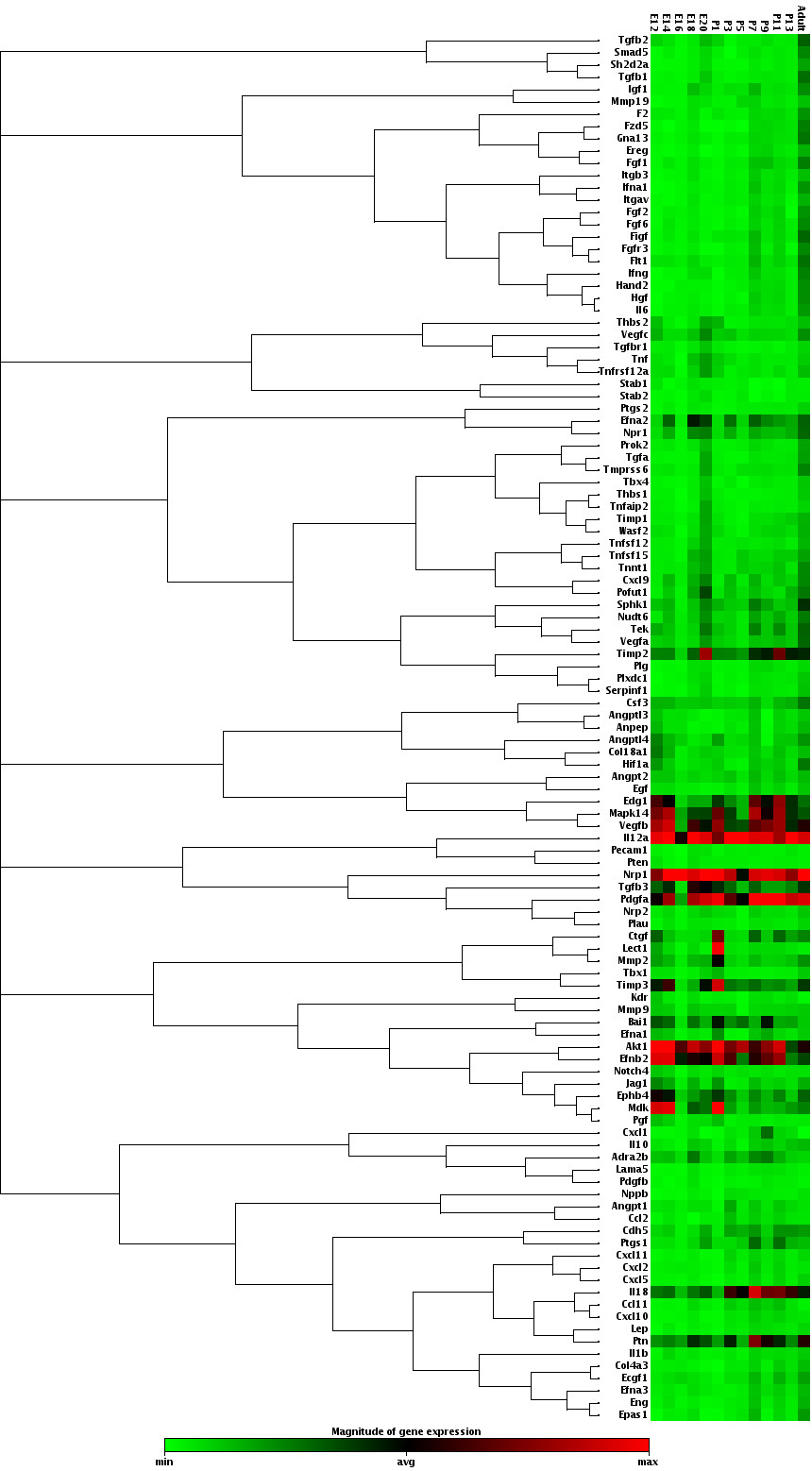
Our array analysis showed that 62 of 113 angiogenesis-related genes are expressed at varying levels during murine brain development (Figure 1, Table 2, Additional file 1: Table S1). The remaining 51 genes are either not expressed at all or expressed by a specific cell population at very minute levels, thus not detected in the array system. We have selected three novel genes, Bai1, Nudt6, and Npr1, to confirm the array data by qPCR analysis and observed similar expression patterns between array and qPCR (Additional file 2: Figure S1). Among these genes, Nudt6 is highly novel and there is no study pinpointing to its expression in any developmental process or stage. Nudt6 is synthesized from the complementary strand of Fgf2, and was implicated to function in post-transcriptional regulation of Fgf2 [24]. Studies on Nudt6 are very limited, and its roles are still unknown [25]. This study demonstrates the expression of Nudt6 during brain development for the first time, and its relatively high expression implies that it might have various crucial roles during CNS development (Table 2).

Figure 1
Figure 1 caption
Clustergram analysis and heatmap graph of gene expression data. Developmental expression level of 113 angiogenesis-related genes was shown as heatmap graph and genes were clustered according to their expression patterns.
Comparative analysis of the array data revealed remarkable results: Between the three isoforms of widely known angiogenic stimulators, Vegf-A, -B and -C, Vegf-B had a 5-fold higher expression than the other two (Table 2), indicating it might have pivotal roles in neurodevelopment in addition to vascular development. Likewise, Nrp1, a co-receptor for Vegf-B and an axon guidance molecule of the semaphorin protein family, was expressed ten-fold more than the other two specific Vegf receptors, Flt1 and Kdr (Table 2). Also, between angiopoietin molecules (Ang1 and Ang2), which play significant roles in endothelial cell lumen stability [1], we found that Ang2 expression was equal to Vegf-A and Vegf-C, but its antagonist Ang1 seem to have no role during brain development (Table 2). Another notable finding is the Ephrin family member, Efnb2, which was 3–10 times more expressed than the other three ephrin ligands, Efna1, 2, 3, throughout brain development (Table 2). Similar comparisons are also possible for other major angiogenic molecules (like Tgf family members, Interleukins, Mmp family etc.) (Table 2). Such comparative analysis, (i.e. to be able to see the expression of all members of the same or close gene families at the same set) might be useful for further functional studies.
Differentially expressed genes between embryonic and postnatal stages
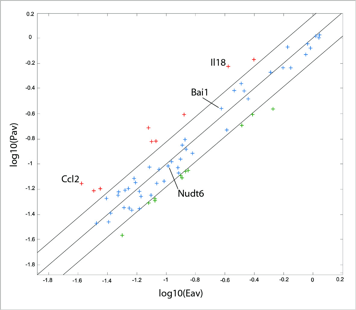
Among the 62 genes that were expressed, 19 displayed more than 1.5 fold higher expression in either stage of the development (Figure 2). Eleven genes [Tgfbr1 (Eav/Pav:1.84), Pgf (1.54), Tnf (1.59), Tnfrsf12a (1.65), Vegfc (1.55), Col18a1 (1.65), Jag1 (1.55), Pofut1 (1.57), Ephb4 (1.60), Tgfb3 (1.55), and Mdk (1.94)] were expressed higher in embryonic stage and eight genes [Ccl2 (Pav/Eav:2.64), Figf (1.91), Fgf1 (1.79), Lect1 (2.57), Ptgs1 (1.88), Cdh5 (1.78), Ctgf (1.88), Il18 (2.23)] displayed higher levels of expression in postnatal stage. Among these, only Ccl2 and Il18 have reached to a statistically significant difference (p: 0.011 and 0.028, respectively).

Figure 2
Figure 2 caption
Scatter plot analysis of array results shows the differentially expressed genes between embryonic and postnatal stages. Graph denotes the logarithmic values of average gene expression in either stage. A total of 11 genes, Tgfbr4, Pgf, Tnf, Tnfrsf12a, Vegfc, Col18a1, Jag1, Pofut1, Ephb4, Tgfb3, and Mdk displayed higher expression in embryonic stage (shown in green). Eight genes, Ccl2, Figf, Fgf1, Lect1, Ptgs1, Cdh5, Ctgf, and Il18 showed a higher level of expression at postnatal stage (shown in red), but with a significant difference for only Ccl2 (p: 0.011) and Il18 (0.028) between two phases. House-keeping genes were equally expressed in both stages, except for the B2m. Boundary value was assigned as 1.5 fold.
Ccl2 (chemokine (C-C motif) ligand 2) has been previously identified as a small cytokine involved in several inflammatory processes, but later it has been proposed to have functions in modulation of neuronal activity and neuroendocrine signaling and it was constitutively expressed by astrocytes [26, 27]. Astrocytes begin to arise at late embryonic stages and populate the cortex largely within the first three weeks of postnatal development [28]. We have found that Cc12 expression increases 2.64-fold during postnatal development (Table 2, Figure 2) overlapping with astrocyte production, which indicates that neuromodulatory function of astrocytes might be mediated through the Ccl2.
Another significant molecule is Il18, previously not implicated in brain development. We have found that Il18 is strongly expressed throughout development with two-fold more expression in postnatal stage (Figure 2, Table 2). This data indicate that, besides its known function as a pro-inflammatory molecule [29], Il18 seem to have important roles in neuronal and vascular development; further functional studies may give new insights into the biology of this molecule. It might be also worthwhile to investigate the tissue level expression of Il18 in cerebrovascular malformations, since Il18 was shown to have a role in pathological angiogenesis in other diseases, like rheumatoid arthritis [30, 31]. Such studies may be especially valuable, since Il18 can be used as a prognostic or clinical marker for cerebrovascular malformations and may be assessed through the analysis of cerebrospinal fluid.
Bai1 is expressed on growing cerebral blood vessels
Bai1 is a recently identified G-protein coupled receptor that has been implicated to have a role in a few physiological and pathological processes. It has been originally discovered as an anti-angiogenic molecule acting under the regulation of p53 in an
In adult mice brain, Bai1 is expressed by astrocytes, by almost all neurons and low levels by macrophages [42, 43]. Based on its regulatory domains and interacting partners, Bai1 was hypothesized to have roles in cell adhesion, growth cone guidance, neurotransmitter release and potentially in some signal transduction pathways [33], however, there is no direct evidence regarding its biological functions in neural tissue.
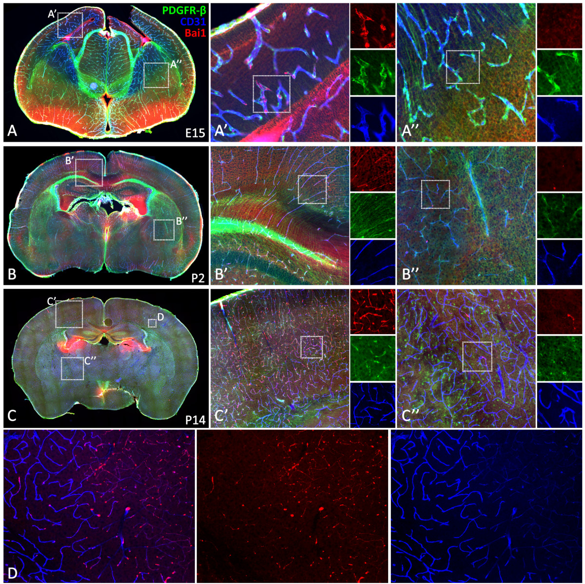
Here, we show that, Bai1 is strongly expressed throughout mouse neurodevelopment at varying levels (Figure 1, Table 2), on average 4- to 5-fold greater than that of Vegf-A receptors, Flt1 and Kdr (Table 2). Our co-immunohistochemistry analysis with endothelial and pericyte cell markers CD31 and PDGFR-beta revealed that, Bai1 is expressed on growing blood vessels of mice brain (Figure 3). Initially around E15, Bai1 is expressed only in the vessels of dorsal cerebral cortex and hippocampus, but not in other brain regions (Figure 3A). At P2, Bai1's expression expands toward the more dorsolateral and ventral cortical areas reaching up to the piriform cortex, but being still absent in subcortical areas (Figure 3B). At P14, Bai1 is strongly expressed in all cortical microvessels as they occupy the whole cortex and to some extent in some of the lateral striatal vessels and dorsal thalamic nuclei, but not in ventral thalamic and hypothalamic vessels at this age (Figure 3C). Notably, expression of the Bai1 was high at the tips of the angiogenic sprouts and branching points, where its function is potentially more needed (Figure 3C’ and D). In those brain parts in which Bai1 is not active, other members of the Bai family, Bai2 and Bai3 might be functioning, especially given the fact that most of the regulatory domains are highly conserved between these members [33].

Figure 3
Figure 3 caption
Bai1 is differentially expressed on growing blood vessels during mice brain development. Tissue-level characterization of Bai1 expression at E15 (A), P2 (B) and P14 (C, D) showed that Bai1 was expressed by cortical blood vessels throughout development (A’, B’ and C’). Initially expressed only by cortical vessels (A’, A”), however, as development progresses, striatal and thalamic vessels also started to express but weakly and non-homogeneously (A”, B”, and C”). Interestingly, at P14, expression was not homogenous in all blood vessels and especially notable on small dense microvessels in the cortex (D).
We also noticed that Bai1 expression is not homogenous throughout the cortex and generally more prominent around dense microvessels at P14 (Figure 3D). This might be because Bai1 function is potentially required especially for endothelial cells found on the angiogenically active small blood vessels to regulate new vessel formation. An alternative reason for this observation might be that the antibody that we used in this study recognizes the N-terminal extracellular domain of the Bai1 (epitope corresponding to amino acids 81–350), which contains a portion of the thrombospondin type-1 repeats found in soluble Vstat40 and Vstat 140 molecules. These thrombospondin type-1 repeats were shown to interact with the CD36 receptor, which is expressed only by endothelial cells on microvessels, but not on larger vessels [33]. Thus, the antibody might be detecting the soluble cleaved product of Bai1 more prominently on small microvessels.
Conclusion
Expression analysis of angiogenesis-related genes in brain development is a critical initial step to elucidate possible mechanisms and genes that are involved in cerebrovascular pathologies. The data presented here revealed novel angiogenesis-related genes, like Bai1 and Nudt6 that play roles in brain development and in this manner, expected to contribute to basic and clinical research on cerebrovascular diseases.
Additional files
Additional file 1: Table S1. Normalized expression levels of genes analyzed on the arrays. (XLS 79 KB)
Additional file 2: Figure S1. qPCR expression analysis of Bai1, Nudt6 and Npr1. Experiments repeated three times, error bars shows the standard errors, GAPDH normalized mean values of the three measurements were used. (TIFF 7 MB)
Acknowledgement
Turker Kilic MD PhD is a member of the Turkish Academy of Sciences (TUBA). This study was supported by TUBITAK (SBAG-108S080), Marmara University Research Funds (SAG-B-030108-0008), Boğaziçi University Research Funds (07HB104), Suna and Inan Kirac Foundation and TUBA.
Authors’ original submitted files for images
Below are the links to the authors’ original submitted files for images.
Authors’ original file for figure 1
Authors’ original file for figure 2
Authors’ original file for figure 3
Authors’ original file for figure 4
Authors’ original file for figure 5
Authors’ original file for figure 6
References
- Angiogenesis: regulators and clinical applications. Biochem Pharmacol. 2001;61:253-270.
- Angiogenesis in cancer and other diseases. Nature. 2000;407:249-257.
- Angiogenesis in life, disease and medicine. Nature. 2005;438:932-936.
- Vascular malformations ("angiomas") of the dura mater. J Neurosurg. 1966;25:309-311.
- Report on the cooperative study of intracranial aneurysms and subarachnoid hemorrhage. IV. Cerebral angiography. An analysis of the diagnostic value and complications of carotid and vertebral angiography in 5,484 patients. J Neurosurg. 1966;25:98-114.
- AVM of the brain: history, embryology, pathological considerations, hemodynamics. Microsurgery. Volume IIIA. 1987.:48-.
- Vascular malformations of the central nervous system. J Neuropathol Exp Neurol. 1995;54:609-621.
- Aberrant angiogenic characteristics of human brain arteriovenous malformation endothelial cells. Neurosurgery. 2009;64:139-146.
- Endothelial cell culture from human cerebral cavernous malformations. Stroke. 1998;29:2426-2434.
- Expression of structural proteins and angiogenic factors in cerebrovascular anomalies. Neurosurgery. 2000;46:1179-1191.
- Evidence of increased endothelial cell turnover in brain arteriovenous malformations. Neurosurgery. 2001;49:124-131.
- Coexpression of angiogenic factors in brain arteriovenous malformations. Neurosurgery. 2005;56:1058-1065.
- Inhibition of angiogenesis induced by cerebral arteriovenous malformations using gamma knife irradiation. J Neurosurg. 2007;106:463-469.
- Endovascular Treatment Increases but Gamma Knife Radiosurgery Decreases Angiogenic Activity of Arteriovenous Malformations: An in Vivo Experimental Study Using a Rat Cornea Model. Neurosurgery. 2010;66:121-130.
- Cerebral arteriovenous malformations. Part 1: cellular and molecular biology. Neurosurg Focus. 2009;26:E10-.
- Cerebral arteriovenous malformations. Part 2: physiology. Neurosurg Focus. 2009;26:E11-.
- Embryological basis of some aspects of cerebral vascular fistulas and malformations. J Neurosurg. 1996;85:1-8.
- Biology of vascular malformations of the brain. Stroke. 2009;40:e694-e702.
- Pathobiology of human cerebrovascular malformations: basic mechanisms and clinical relevance. Neurosurgery. 2004;55:1-16.
- Increased expression of ephrin A1 in brain arteriovenous malformation: DNA microarray analysis. Neurosurg Rev. 2007;30:299-305.
- Expression of integrins in cerebral arteriovenous and cavernous malformations. Neurosurgery. 2006;58:159-168.
- The genetics of cerebrovascular malformations. J Stroke Cerebrovasc Dis. 2002;11:279-287.
- Endothelial expression of constitutively active Notch4 elicits reversible arteriovenous malformations in adult mice. Proc Natl Acad Sci U S A. 2005;102:9884-9889.
- The fibroblast growth factor-2 antisense gene inhibits nuclear accumulation of FGF-2 and delays cell cycle progression in C6 glioma cells. Mol Cell Endocrinol. 2007;267:127-136.
- Characterization and tissue-specific expression of the rat basic fibroblast growth factor antisense mRNA and protein. Proc Natl Acad Sci U S A. 1997;94:4943-4947.
- Human monocyte chemoattractant protein-1 (MCP-1). Full-length cDNA cloning, expression in mitogen-stimulated blood mononuclear leukocytes, and sequence similarity to mouse competence gene JE. FEBS Lett. 1989;244:487-493.
- Highly regionalized neuronal expression of monocyte chemoattractant protein-1 (MCP-1/CCL2) in rat brain: evidence for its colocalization with neurotransmitters and neuropeptides. J Comp Neurol. 2005;489:275-292.
- Ge WP, Miyawaki A, Gage FH, Jan YN, Jan LY: Local generation of glia is a major astrocyte source in postnatal cortex. Nature. , 484: 376-380.
- Interleukin-18 binding protein: a novel modulator of the Th1 cytokine response. Immunity. 1999;10:127-136.
- Interleukin 18 induces angiogenesis in vitro and in vivo via Src and Jnk kinases. Ann Rheum Dis. 2010;69:2204-2212.
- Evidence of IL-18 as a novel angiogenic mediator. J Immunol. 2001;167:1644-1653.
- A novel brain-specific p53-target gene, BAI1, containing thrombospondin type 1 repeats inhibits experimental angiogenesis. Oncogene. 1997;15:2145-2150.
- Emerging roles for the BAI1 protein family in the regulation of phagocytosis, synaptogenesis, neurovasculature, and tumor development. J Mol Med (Berl). 2011;89:743-752.
- Vasculostatin, a proteolytic fragment of brain angiogenesis inhibitor 1, is an antiangiogenic and antitumorigenic factor. Oncogene. 2005;24:3632-3642.
- Vasculostatin inhibits intracranial glioma growth and negatively regulates in vivo angiogenesis through a CD36-dependent mechanism. Cancer Res. 2009;69:1212-1220.
- A proprotein convertase/MMP-14 proteolytic cascade releases a novel 40 kDa vasculostatin from tumor suppressor BAI1. Oncogene. 2012.
- Diverse somatic mutation patterns and pathway alterations in human cancers. Nature. 2010;466:869-873.
- Brain angiogenesis inhibitor 1 is differentially expressed in normal brain and glioblastoma independently of p53 expression. Am J Pathol. 2003;162:19-27.
- Brain-specific angiogenesis inhibitor 1 expression is inversely correlated with vascularity and distant metastasis of colorectal cancer. Int J Oncol. 1998;13:967-970.
- Expression of VEGF and brain specific angiogenesis inhibitor-1 in glioblastoma: prognostic significance. Oncol Rep. 2004;11:863-869.
- BAI1 is an engulfment receptor for apoptotic cells upstream of the ELMO/Dock180/Rac module. Nature. 2007;450:430-434.
- Brain-specific angiogenesis inhibitor-1 expression in astrocytes and neurons: implications for its dual function as an apoptotic engulfment receptor. Brain Behav Immun. 2011;25:915-921.
- Characterization of mouse brain-specific angiogenesis inhibitor 1 (BAI1) and phytanoyl-CoA alpha-hydroxylase-associated protein 1, a novel BAI1-binding protein. Brain Res Mol Brain Res. 2001;87:223-237.

