
Human and mouse brain-derived endothelial cells require high levels of growth factors medium for their isolation, in vitro maintenance and survival
Article Sidebar
Published
May 14, 2013
Main Article Content
Abstract
Background
Brain microvascular endothelial cells (BMVECs) constitute the primary limitation for passage of ions and molecules from the blood into the brain through the blood brain barrier. Numerous multi-step procedures for isolating and culturing BMVECs have been described. However, each one demonstrates major limitations in purity of culture and/or low proliferation rate. Our goal was to study the efficiency of our pending patent medium, Endothelial Proliferation Medium (EndoPM), on the isolation and purification of human and murine BMVECs.Methods
BMVECs, cultured in EndoPM were compared to those cultured in a commercial medium EBM. Cultures were characterized by flow cytometric analysis, lineage differentiation, the ability to form tube-like structure, immunofluorescence, molecular analyses and also in an in vivomodel assay. Moreover permeability was assayed by monitoring the passage of Dextran-FITC through a tight monolayer of BMVECs grown to confluence in Boyden chambers. One way Anova two-tailed test was utilized for all statistical analyses.
Results
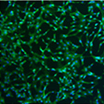
The properties of ECs in human and murine BMVECs is confirmed by the expression of endothelial markers (CD31, CD105, CD146, Tie-2 and vWF), of representative proangiogenic genes (ICAM1, VCAM1 and integrin ITGAV), of considerable tube-forming ability, with low-density lipoprotein uptake, eNOS and GLUT-1 expression. Furthermore cells are able to express markers of the junctional architecture as VE-cadherin, β-catenin and Claudin-5 and greatly reduce dextran permeability as barrier functional test. Moreover BMVECs spontaneously organize in vascular-like structures and maintain the expression of endothelial markers in an in vivoxenograft model assay. The significant effect of EndoPM is confirmed by the study of proliferation index, survival index and the behaviour of BMVECs and fibroblasts in co-culture conditions.
Conclusion
Herein we describe a simple and reproducible method for the isolation and expansion of human and mouse BMVECs, based on a newly formulated medium (EndoPM) with optimized concentration of growth factors (EGF, FGF-2 and Bovine Brain Extract-BBE). This procedure should facilitate the isolation and expansion of human and mouse BMVECs with extended lifetime, good viability and purity. This approach may provide an effective strategy to aid phenotypical and functional studies of brain vessels under physiological and pathological conditions.Article Details
How to Cite
NAVONE, Stefania Elena et al.
Human and mouse brain-derived endothelial cells require high levels of growth factors medium for their isolation, in vitro maintenance and survival.
Vascular Cell, [S.l.], v. 5, n. 1, p. 10, may 2013.
ISSN 2045-824X.
Available at: <https://vascularcell.com/index.php/vc/article/view/10.1186-2045-824X-5-10>. Date accessed: 21 july 2026.
doi: http://dx.doi.org/10.1186/2045-824X-5-10.
Section
Original Research