The role of angiogenesis in the pathology of multiple sclerosis
Main Article Content
Abstract
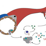
Angiogenesis, or the growth of new blood vessels from existing vasculature, is critical for the proper development of many organs. This process is inhibited and tightly regulated in adults, once endothelial cells have acquired organ-specific properties. Within the central nervous system (CNS), angiogenesis and acquisition of blood–brain barrier (BBB) properties by endothelial cells is essential for CNS function. However, the role of angiogenesis in CNS pathologies associated with impaired barrier function remains unclear. Although vessel abnormalities characterized by abnormal barrier function are well documented in multiple sclerosis (MS), a demyelinating disease of the CNS resulting from an immune cell attack on oligodendrocytes, histological analysis of human MS samples has shown that angiogenesis is prevalent in and around the demyelinating plaques. Experiments using an animal model that mimics several features of human MS, Experimental Autoimmune Encephalomyelitis (EAE), have confirmed these human pathological findings and shed new light on the contribution of pre-symptomatic angiogenesis to disease progression. The CNS-infiltrating inflammatory cells that are a hallmark of both MS and EAE secrete several factors that not only contribute to exacerbating the inflammatory process but also promote and stimulate angiogenesis. Moreover, chemical or biological inhibitors that directly or indirectly block angiogenesis provide clinical benefits for disease progression. While the precise mechanism of action for these inhibitors is unknown, preventing pathological angiogenesis during EAE progression holds great promise for developing effective treatment strategies for human MS.
Article Details
How to Cite
LENGFELD, Justin; CUTFORTH, Tyler; AGALLIU, Dritan.
The role of angiogenesis in the pathology of multiple sclerosis.
Vascular Cell, [S.l.], v. 6, n. 1, p. 23, nov. 2014.
ISSN 2045-824X.
Available at: <https://vascularcell.com/index.php/vc/article/view/10.1186-s13221-014-0023-6>. Date accessed: 21 feb. 2026.
doi: http://dx.doi.org/10.1186/s13221-014-0023-6.
Section
Review

