
Positron emission tomography detection of human endothelial cell and fibroblast monolayers: effect of pretreament and cell density on 18FDG uptake
Article Sidebar
Published
Mar 20, 2012
Main Article Content
Abstract
Background
The non-destructive assessment and characterization of tridimensional (3D) cell and tissue constructs in bioreactors represents a challenge in tissue engineering. Medical imaging modalities, which can provide information on the structure and function of internal organs and tissues in living organisms, have the potential of allowing repetitive monitoring of these 3D cultures in vitro. Positron emission tomography (PET) is the most sensitive non-invasive imaging modality, capable of measuring picomolar amounts of radiolabeled molecules. However, since PET imaging protocols have been designed almost exclusively for in vivoinvestigations, suitable methods must be devised to enable imaging of cells or tissue substitutes. As a prior step to imaging 3D cultures, cell radiotracer uptake conditions must first be optimized.
Methods
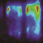
In this study, human umbilical vein endothelial cells (HUVEC) and human fibroblasts were cultured at different densities and PET was used to non-destructively monitor their glycolytic activity by measuring 18F-fluorodeoxyglucose ( 18FDG) uptake. Various cell preconditioning protocols were investigated by adjusting the following parameters to optimize 18FDG uptake: glucose starvation, insulin stimulation, glucose concentration, 18FDG incubation time, cell density and radiotracer efflux prevention.
Results
The conditions yielding optimal 18FDG uptake, and hence best detection sensitivity by PET, were as follows: 2-hour cell preconditioning by glucose deprivation with 1-hour insulin stimulation, followed by 1-hour 18FDG incubation and 15-minute stabilization in standard culture medium, prior to rinsing and PET scanning.
Conclusions
A step-wise dependence of 18FDG uptake on glucose concentration was found, but a linear correlation between PET signal and cell density was observed. Detection thresholds of 36 ± 7 and 21 ± 4 cells were estimated for endothelial cells and fibroblasts, respectively.
Article Details
How to Cite
CHOUINARD, Julie A et al.
Positron emission tomography detection of human endothelial cell and fibroblast monolayers: effect of pretreament and cell density on 18FDG uptake.
Vascular Cell, [S.l.], v. 4, n. 1, p. 5, mar. 2012.
ISSN 2045-824X.
Available at: <https://vascularcell.com/index.php/vc/article/view/10.1186-2045-824X-4-5>. Date accessed: 21 july 2026.
doi: http://dx.doi.org/10.1186/2045-824X-4-5.
Section
Original Research